Molecular Biology Group
Functional Analysis of the Multifunctional Protein Praja1
Overview
In the Molecular Biology Group, we conduct research aimed at understanding life at the molecular level through the functional analysis of proteins, the cellular workers. Currently, our focus is on a protein called Praja1, possessing two distinct molecular functions.
Praja1 functions both as an “E3 ubiquitin ligase,” which recognizes and directs the degradation of unwanted proteins, and as a “DNA-protective protein” that safeguards nuclear DNA from damage. Our research comprehensively examines both of these aspects.
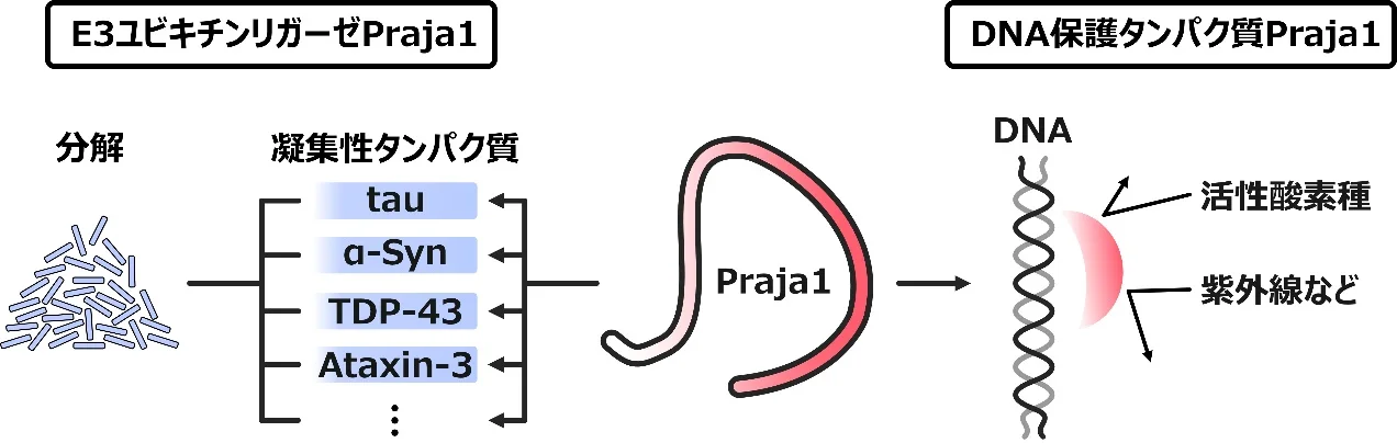
Praja1 as an “E3 ubiquitin ligase”
Protein expression in living organisms is strictly regulated in terms of when, where, and how much is expressed. Among the enzymes involved in this process, E3 ubiquitin ligases (E3 enzymes) are known to direct the degradation of proteins that are no longer needed. Inadequate E3 enzyme activity is thought to lead to the accumulation of toxic protein waste, which in turn links to disease onset and aging.
Since Praja1 broadly recognizes and degrades aggregating proteins commonly found in neurodegenerative diseases (such as α-synuclein, TDP-43, and FUS), it is suggested to play a suppressive role in the onset and progression of these diseases. Our research has revealed that Praja1 also recognizes and degrades Tau protein, which aggregates in neurodegenerative diseases such as Alzheimer’s disease1. We are currently exploring the interaction between Praja1 and Tau in greater depth. Additionally, collaborating with the electrochemistry group within our laboratory, we are developing biosensors utilizing Praja12.
Praja1 as a “DNA-protective protein”
Intracellular DNA is damaged daily by various factors. In addition to internal factors such as reactive oxygen species generated by metabolic activity within the body, it is also exposed to external factors such as ultraviolet radiation and radiation therapy for cancer. Furthermore, the cosmic rays that humans are exposed to during space travel can damage DNA, which will pose a challenge in the future as humanity seeks to improve its quality of life in space (space QOL).
Through molecular evolutionary analysis, we discovered that Praja1 acquired the ability to localize to the cell nucleus during the evolution of mammals3. Furthermore, while investigating the physiological role of Praja1 after its entry into the nucleus, we obtained a novel finding—previously unreported—that Praja1 protects DNA4. We are currently working to elucidate the molecular mechanisms by which Praja1 exerts its DNA-protective effects.
Related Publications
- [1] Shiho Aoki✞, Wataru Onodera✞,※, Akihiko Takashima, Kotaro Kawasaki, Kazuki Imadegawa, Hikaru Kurahashi, Mizuho Oishi, Toru Asahi, Yoshiyuki Soeda. E3 ligase Praja1 mediates ubiquitination and degradation of microtubule-associated protein tau. The FEBS Journal. 293(8): 2212-2224. (2025)
✞ Shiho Aoki and Wataru Onodera equally contributed to this work.
※ Corresponding author
This paper was selected as an Editor’s Choice article. - [2] Wataru Mori, Wataru Onodera, Terutoshi Kojima, Toru Asahi※, Takuya Nakanishi※. Interaction between intrinsically disordered proteins Praja1 and α-synuclein on gold electrodes. Chemistry Letters. 53(11):upae205. (2024)
※ Corresponding author - [3] Wataru Onodera✞,※, Kotaro Kawasaki✞, Mizuho Oishi, Shiho Aoki, Toru Asahi※. Functional Divergence and Origin of the Vertebrate Praja Family. Journal of Molecular Evolution. 92:21-29. (2024)
✞ Wataru Onodera and Kotaro Kawasaki contributed equally to this work.
※ Corresponding author - [4] Kotaro Kawasaki, Toru Asahi, Wataru Onodera※. Praja1 protects cells from DNA damage through direct DNA binding. bioRxiv. https://doi.org/10.64898/2025.12.04.691747 (2025)
※ Corresponding author
Insect Group
Analysis of Social Behavior Using Deep Learning
Overview
The Insect Group is working to elucidate the molecular basis of social behavior using crickets as a model organism. Specifically, we use deep learning to track courtship and fighting behaviors with high precision, visualizing detailed behavioral patterns. In addition, by integrating behavioral data with omics data, we aim for a comprehensive understanding of social behavior. This research is a collaboration with the Suzuki Lab (https://web.tuat.ac.jp/~tszk/index.html) and the Kataoka Lab (https://web.tuat.ac.jp/~ttanaka/research/bio-info/03.html) at the Tokyo University of Agriculture and Technology.
For more details on the research and achievements, please see: https://web.tuat.ac.jp/~ttanaka/research/bio-info/03.html
Health Science Group
What does it mean, SCIENTIFICALLY, for something to be "good for the body"?
In the Health Science Group, we take the question of what it really means for something to be "good for the body" as our keyword, working to clarify, at both the whole-body and molecular levels, how naturally derived bioactive compounds affect a range of health problems and diseases. In particular, we focus on chronic inflammation, held to lie at the root of nearly all chronic diseases, and use animal models and cultured cells to comprehensively study how the things we eat each day act on the body's internal systems and what results from that in the body as a whole.
The two faces of endocannabinoids in obesity-driven systemic inflammation
Overeating and obesity trigger inflammation throughout the body, and this is known to underlie a range of disorders, from diabetes to chronic pain. We focus on the endocannabinoid system, particularly the CB2 receptor expressed on immune cells, which is known as one of the regulatory systems for systemic inflammation.
In nerve injury, the CB2 receptor suppresses inflammation and pain. In inflammation associated with diet-induced obesity, however, we found the opposite: it drives inflammation and pain in peripheral nerves (Hosoki et al., Life Sciences, 2024). We have further found that this switch between "suppressing" and "promoting" is closely tied to age: even on the same diet, the way inflammation-driving immune cells accumulate changes markedly at a relatively early stage, corresponding roughly to the teens through thirties in humans. We are now working to uncover, at the molecular level, why the CB2 receptor shows such two faces, and how the immune cells behind them are generated and recruited.
Approaching sex differences in metabolism through an edible-insect factor that fattens only males
Even when we eat the same thing, the body responds differently from person to person, and between the sexes. Metabolism in particular shows marked sex differences: it is men who tend to accumulate visceral fat and to suffer the more serious consequences of obesity. Yet why men are especially prone to weight gain remains almost entirely unexplained at the level of the body's mechanisms.
We found that when a certain edible insect powder is mixed in small amounts into a standard diet, male mice alone gain weight markedly, even though food intake and caloric intake are unchanged. Females do not gain weight at all, and this difference cannot be explained by levels of sex hormones alone. It points to a male-specific metabolic mechanism that does not rely on sex hormones. We are now working to identify the active component derived from this insect, and to pin down where in the body it acts to fatten only males, closing in on the metabolic core that gives rise to this sex difference.
How everyday components of the diet relate to inflammation and the body
What we examine in asking "what is good for the body" is not only the special compounds. We also pursue how familiar, everyday components of the diet affect the body. For example, we have reported that dietary fiber, generally regarded as "good for you," can in fact have adverse effects unless the type is chosen with care (Ito et al., ACS Pharmacology and Translational Science, 2024). Beyond this: how excess salt alters the course of obesity-driven inflammation and pain; how components of spices engage in the regulation of inflammation; and how, as in food allergy, the immune response to what we eat ripples through the body. We take up these diverse questions through a single lens: the relationship between what we eat each day and how the body responds.
Computational Biology Group
Elucidating Tissue 3D Architecture and Brain Vascular Aging with Public Data
Overview
In the Computational Biology Group, we use computational science to read out the rapidly expanding body of public gene-expression data and to reveal, at scale, how tissues are organized and how they are remodeled with aging and disease. Our particular focus is developing analysis tools for spatial transcriptomics, which measures where in a tissue gene expression occurs while preserving location, and connecting this to the understanding of tissues such as the brain vasculature. We pursue this question with two wheels turning together, large-scale dry analysis and experimental (wet) validation through collaboration, and we also develop the computational tools this requires (Figure 1).
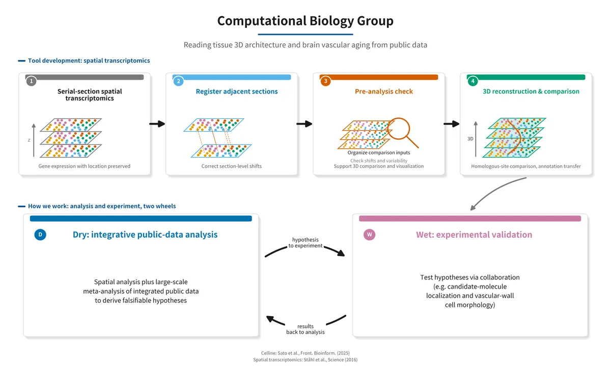
Figure 1. Overview of the Computational Biology Group. We develop a tool that aligns serial-section spatial transcriptomics for comparing and visualizing tissue 3D architecture. Around this, we support 3D reconstruction, homologous-site comparison, and annotation transfer. Together with large-scale meta-analysis built on public-data integration foundations, falsifiable hypotheses from dry analysis are tested by experiment (wet) through collaboration, and the results feed back into the analysis, forming a two-wheel cycle.
Reading tissue 3D architecture from spatial transcriptomics
Spatial transcriptomics measures where in a tissue gene expression occurs while preserving location, and it has been transforming the life sciences in recent years [1]. However, a single experiment usually yields one thin section (a 2D slice). To study the 3D architecture that the tissue actually has, or to compare the same anatomical region across samples, serial sections must be organized computationally while accounting for section-level shifts and data variability.
The tool we are currently developing is a computational foundation for comparing and visualizing tissue 3D structure from serial sections. It organizes the data into a form that supports 3D reconstruction, homologous-site comparison, and annotation transfer while taking section-level shifts and variability into account. We are currently checking its behavior on public datasets and organizing the evaluation for broader applications.
Foundations for integrating public data
Large-scale meta-analysis requires a foundation for retrieving, standardizing, and integrating public data scattered across repositories such as GEO and SRA. Within the group, we have built public-data integration foundations such as Celline [5], which executes this whole sequence with a single-line command. With these, we can, for example, integrate many public human-brain single-nucleus RNA-seq datasets and build cell-type-specific indices of aging. Combined with spatial-transcriptomics analysis, this lets us approach how the brain vasculature changes along both its 3D architecture and its aging.
Dry and wet: two wheels turning together
What computational analysis provides is, in the end, a falsifiable hypothesis. We therefore emphasize a two-wheel system in which these hypotheses are tested through collaboration with experimental groups. For example, by analyzing public vascular data across several species, we extract candidate molecules and phenotypes related to endothelial and vascular-wall cells. We are now proposing selected candidates to vascular-biology collaborators as experimental protocols to test localization and function. Likewise, we are looking ahead to experimental validation and collaboration on age-related changes in vascular-supporting cells. By confirming computational predictions through experiment and feeding the results back into analysis, we aim to depict how the brain vasculature and other tissues age and progress toward disease.
What we aim for
Developing analysis tools that read tissue structure and change from public data, including spatial transcriptomics, performing large-scale analysis with them, and validating predictions experimentally: by uniting these three elements, we aim to clarify how the brain vasculature changes along both its 3D architecture and its aging, and to openly share the tools we develop with the community. This is the goal of the Computational Biology Group.
References
- [1] Ståhl PL, et al. Visualization and analysis of gene expression in tissue sections by spatial transcriptomics. Science 353: 78–82 (2016).
- [2] Vanlandewijck M, et al. A molecular atlas of cell types and zonation in the brain vasculature. Nature 554: 475–480 (2018).
- [3] Yang AC, et al. A human brain vascular atlas reveals diverse mediators of Alzheimer's risk. Nature 603: 885–892 (2022).
- [4] Montagne A, et al. Blood-brain barrier breakdown in the aging human hippocampus. Neuron 85: 296–302 (2015).
Related Publications
- [5] Sato Y, Asahi T, Kataoka K. Celline: a flexible tool for one-step retrieval and integrative analysis of public single-cell RNA sequencing data. Front. Bioinform. 5: 1684227 (2025).
Cultivated Meat Group
A circular cell culture system using microalgae and mammalian cells for the production of sustainable cultured meat
The cultivated meat group is conducting research at Tokyo Women’s Medical University, in collaboration with Waseda University, as part of the Moonshot Research and Development Program funded by Japan’s Ministry of Agriculture, Forestry and Fisheries. Our research focuses on developing a sustainable cultivated meat production system known as the Circular Cell Culture (CCC) system, in which photosynthetic algae are utilized to support the growth of animal cells, while spent culture medium from animal cell cultivation is recycled for algal cultivation. Furthermore, the expanded animal cells are assembled into cultivated meat using three-dimensional tissue engineering technologies derived from regenerative medicine. Through the implementation and widespread adoption of this novel circular food production system, we aim to achieve both sustainable food production and environmental conservation.
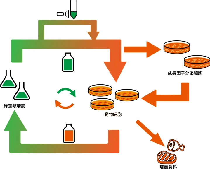
Figure 1. A grapical abstract of circular cell culture system
Algae as an Alternative Source of Culture Medium Nutrients
To cultivate mammalian cells in vitro, nutrients such as carbohydrates, nitrogen sources, and vitamins are required to support cell survival and proliferation. Conventional basal media used for animal cell culture contain nutrients derived from agricultural crops. However, crop-dependent cell culture may contribute to competition among food, livestock feed, and biofuel production, potentially increasing crop prices. In addition, crop production requires extensive land use, water consumption, and the application of fertilizers and pesticides, resulting in significant environmental burdens.
To address these challenges, we have explored the use of algal biomass as an alternative nutrient source. We demonstrated that nutrients essential for animal cell culture, including glucose and amino acids, can be extracted from algae through hydrolysis using ultrasonic treatment, conventional liquid acids such as hydrochloric acid, and environmentally friendly solid acids¹˒². Furthermore, by combining these methods with additional processing techniques, we significantly improved the recovery of amino acids that were difficult to extract using conventional approaches alone. Through optimization of these extraction methods, we achieved algal biomass degradation rates exceeding 90%.
Algal Cultivation Using Spent Animal Cell Culture Medium
Animal cells consume glucose and amino acids as nutrients and produce lactate and ammonia as metabolic waste products. During cell culture, nutrient depletion and waste accumulation inhibit cell survival and proliferation, necessitating periodic replacement of the culture medium.
Spent culture medium still contains residual phosphorus (P), minerals, and ammonia that can serve as nutrients for algal growth. Therefore, it is possible to cultivate algae using waste medium generated from animal cell cultures. Algae naturally assimilate ammonia and convert it into amino acids. In addition, we employ genetically engineered algae capable of converting lactate into pyruvate, a metabolite that can be utilized by animal cells as an energy source³. These engineered algae not only grow using components present in the spent medium but also contribute to its purification.
We confirmed that cultivation of algae in spent animal cell culture medium removed 80–90% of accumulated metabolic waste products. Furthermore, we demonstrated that regenerated culture medium produced through algal upcycling of waste metabolites could support animal cell proliferation⁴. These findings highlight the potential for reducing agricultural resource dependence and minimizing culture waste generation. Collectively, our results demonstrate the feasibility of Cell Culture Circularity (CCC), a system that enables the circular reuse of crop-free culture media.
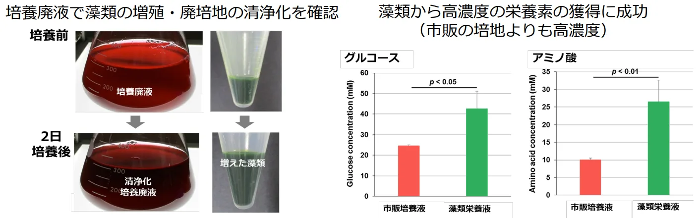
Figure 2. Research progress of circular cell culture system
Production of Cultivated Meat Using Cell Sheet Technology
Cultivated meat is a food product produced by constructing three-dimensional tissues from animal-derived skeletal muscle and adipose cells expanded in vitro using various tissue engineering technologies. As global demand for meat continues to increase alongside population growth, conventional livestock production faces challenges related to infectious disease risks and environmental impacts, including greenhouse gas emissions. Consequently, there is growing interest worldwide in developing cultivated meat production systems that can reduce disease risks through sterile manufacturing processes while minimizing environmental burdens.
A patented technology developed at Tokyo Women’s Medical University utilizes temperature-responsive culture dishes, enabling cultured cells to be harvested as intact sheet-like tissues without enzymatic treatment⁵. These tissue constructs, known as cell sheets, can be layered and fused together to form thicker three-dimensional tissues⁵.
Using this technology, we are developing cultivated chicken meat by stacking cell sheets generated from extensively expanded chicken-derived cells. In addition, we are exploring a variety of tissue engineering approaches—including enzyme-induced gelation, spheroid-based assembly, and cell fiber technologies—to recreate the structure, texture, and overall sensory properties of conventional meat. Through these efforts, we aim to develop cultivated meat products that resemble traditional meat in both form and eating quality.
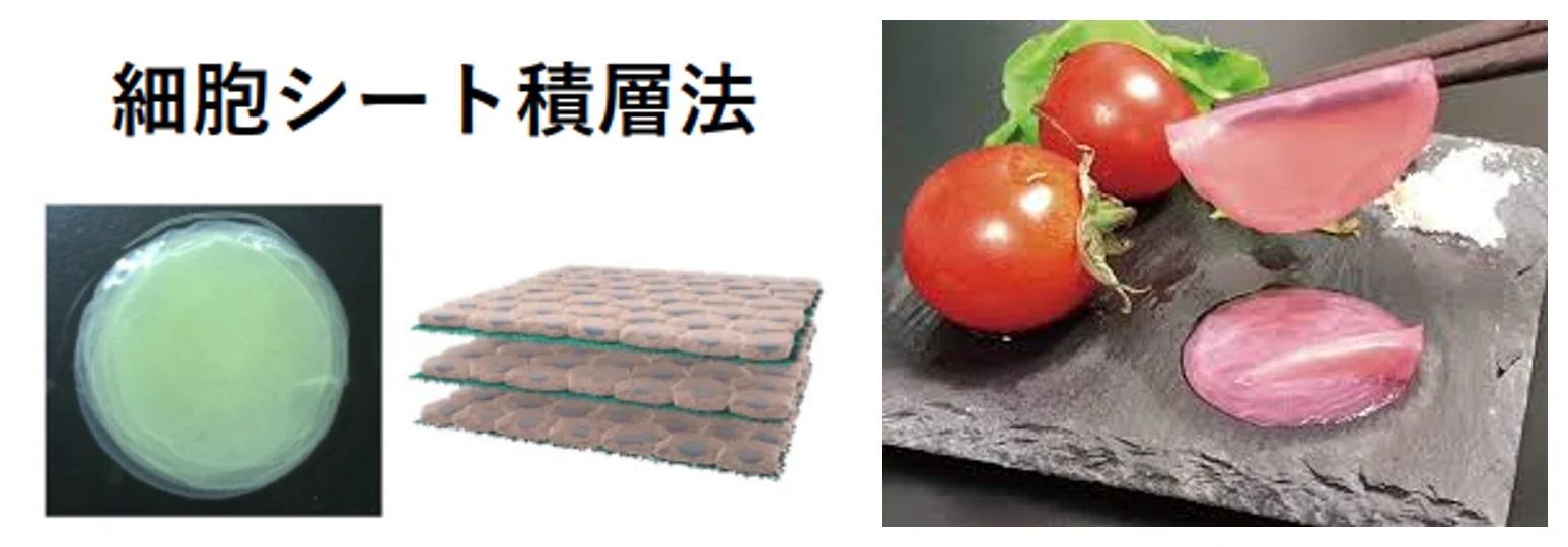
Figure 3. Cell-sheet-based tissue engineering (left) and
cultivated meat produced by stacking 10 cell sheets (right)
References
- [1] Okamoto, Y., Haraguchi, Y., Yoshida, A., Takahashi, H., Yamanaka, K., Sawamura, N., ... & Shimizu, T. (2022). Proliferation and differentiation of primary bovine myoblasts using Chlorella vulgaris extract for sustainable production of cultured meat. Biotechnology Progress, 38(3), e3239.
- [2] Haraguchi, Y., Kato, Y., Inabe, K., Kondo, A., Hasunuma, T., & Shimizu, T. (2023). Circular cell culture for sustainable food production using recombinant lactate-assimilating cyanobacteria that supplies pyruvate and amino acids. Archives of Microbiology, 205(7), 266.
- [3] Shimizu, T., Yamato, M., Kikuchi, A., & Okano, T. (2001). Two-dimensional manipulation of cardiac myocyte sheets utilizing temperature-responsive culture dishes augments the pulsatile amplitude. Tissue engineering, 7(2), 141-151.
Related Publications
- [4] Ghosh, J., Haraguchi, Y., Asahi, T., Nakao, Y., & Shimizu, T. (2023). Muscle cell proliferation using water-soluble extract from nitrogen-fixing cyanobacteria Anabaena sp. PCC 7120 for sustainable cultured meat production. Biochemical and Biophysical Research Communications, 682, 316-324.
- [5] Chu, S., Haraguchi, Y., Asahi, T., Kato, Y., Kondo, A., Hasunuma, T., & Shimizu, T. (2024). A serum-free culture medium production system by co-culture combining growth factor-secreting cells and L-lactate-assimilating cyanobacteria for sustainable cultured meat production. Scientific reports, 14(1), 19578.